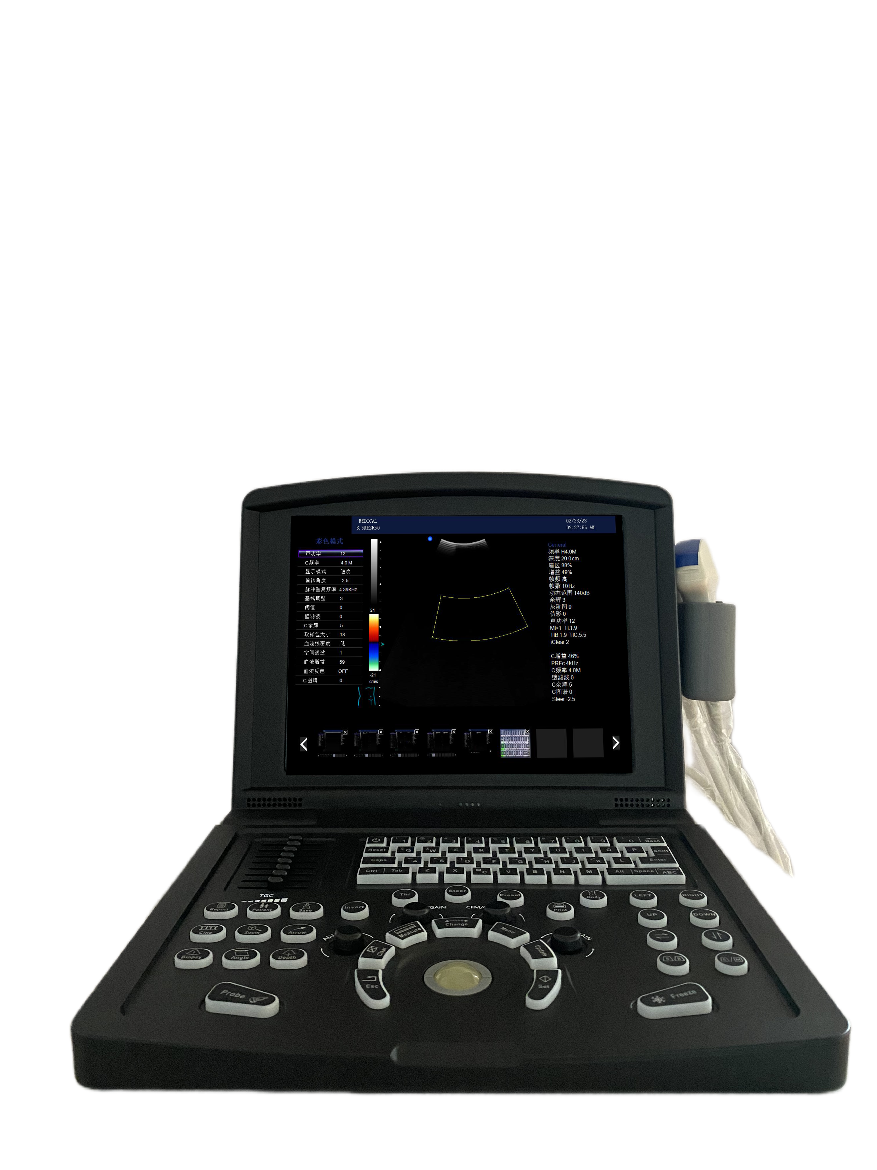
Scanner ad ultrasuoni Doppler a colori portatili per vascolare
$2100-3500 /Set/Sets
| Tipo di pagamento: | T/T |
| Incoterm: | FOB |
| Quantità di ordine minimo: | 5 Set/Sets |
| Trasporti: | Ocean,Land,Air,Express |
| Porta: | Shenzhen,Ningbo,Shanghai |
$2100-3500 /Set/Sets
| Tipo di pagamento: | T/T |
| Incoterm: | FOB |
| Quantità di ordine minimo: | 5 Set/Sets |
| Trasporti: | Ocean,Land,Air,Express |
| Porta: | Shenzhen,Ningbo,Shanghai |
Modello: MDK-680
marchio: Ultrasuoni uniti
Luogo D'origine: Cina
Tipo Di Potere: Elettricità
Servizio Di Garanzia: 1 anno
Assistenza Post-vendita: Pezzi di ricambio gratuiti, Supporto tecnico in linea
Materiale: metallo, plastica
Classificazione Dei Dispositivi Medici: Classe II
Color: white
marchio: United Ultrasound
Monitor Size: 12 inch
Gray Scale: 256 levels
Depth: ≥300mm
Channels: 32
Operating System: Windows 7
| Unità vendibili | : | Set/Sets |
| Tipo pacchetto | : | Cartone |
The file is encrypted. Please fill in the following information to continue accessing it
Scanner ad ultrasuoni Doppler a colori portatili per vascolare (MDK-680)
L'ecografia Doppler a colori vascolari è un metodo di esame ampiamente usato nella nostra vita quotidiana. Questo metodo di esame può verificare principalmente se esiste una trombosi nel corpo del paziente o se c'è malattia nell'arteria del paziente. Può anche verificare se l'arteria del paziente è bloccata.
L'ecografia Doppler a colori può rilevare se vi è placca nell'arteria e se la placca provoca stenosi o occlusione. L'ecografia Doppler di colore vascolare è ampiamente utilizzata nella pratica clinica, utilizzata principalmente per rilevare le seguenti malattie: in primo luogo, se esiste una malattia del sistema venoso. Se c'è trombosi nella vena, se esiste una condizione della valvola, se vi è insufficienza. In secondo luogo, controlla le arterie per la malattia. Come arteriosclerosi, trombosi, stenosi e così via.
I nostri prodotti:
Scanner ad ultrasuoni in bianco e nero
Scanner ad ultrasuoni Doppler a colori
Scanner ad ultrasuoni veterinari


|
No. |
Item |
Index |
|
<1> |
Depth |
≥300mm |
|
<2> |
Lateral Resolution |
≤1mm (Depth≤80mm) ≤2mm (80< Depth≤130mm) |
|
<3> |
Axial Resolution |
≤1mm (Depth≤80mm) ≤2mm (80< Depth≤130mm) |
|
<4> |
Blind Area |
≤4 mm |
|
<5> |
Geometry Position Precision |
horizontal≤5% vertical≤5% |
|
<6> |
Language |
English/Chinese |
|
<7> |
Channels |
32 |
|
<8> |
Displayer |
12” LED |
|
<9> |
External Display |
PAL, VGA, USB |
|
<10> |
Gray Scale |
256 levels |
|
<11> |
Voltage |
AC220V ±10% |
|
<12> |
Operating System |
Windows 7 |
|
<13> |
Scanning Mode |
B, B/B, 4B, B/M, M, B+C, B+D, B+C+D, PDI, CF, PW |
|
<14> |
Probe |
Probe sockets: 2 Probe frequency: 2.0MHz ~ 10.0MHz, 8-step frequency conversion |
|
<15> |
Adjustment parameters of color blood flow image |
Doppler frequency, sampling frame position and size, baseline, color gain, deflection angle, wall filtering, cumulative times, etc |
|
<16> |
Signal processing
|
With dynamic filtering and quadrature demodulation With total gain adjustment Gain adjustment: 8-segment TGC The total gain of Type B, Type C and Type D can be adjusted respectively B/W image gain and color blood flow gain are adjustable respectively |
|
<17> |
Doppler |
Doppler baseline adjustment level 6 Pulse repetition frequency can be adjusted separately: CFM PWD With D linear speed regulation |
|
<18> |
Digital beam forming |
Continuous dynamic focusing of digital beam forming image Full range dynamic aperture of image Dynamic tracing of the whole image Weighted Sum of Image Whole Process Receiving Delay Support half step scanning and ± 10 ° linear receiving deflection angle Multi beam parallel processing technology |
|
<19> |
Basic measurement and calculation function |
Basic measurement in mode B: distance, angle, perimeter and area, volume, stenosis rate, histogram, cross-section |
|
Basic measurement of M-mode: heart rate, time, distance and speed |
||
|
Doppler measurement: time, heart rate, speed, acceleration |
||
|
<20> |
Gynecological measurement and calculation function |
Measurement and calculation of uterus, left ovary, right ovary, left follicle, right follicle, etc |
|
<21> |
Obstetric measurement and calculation function |
G.A, EDD, BPD-FW, FL, AC, HC, CRL, AD, GS, LMP,HL,LV,OFD |
|
<22> |
Urology measurement and calculation function
|
Measurement and calculation of left kidney, right kidney, bladder, residual urine volume, prostate, prostate specific antigen predicted value PPSA, prostate specific antigen density PSAD, etc |
|
<23> |
Product Size |
289×304×222mm |
|
<24> |
Carton Size |
395×300×410mm |
|
<25> |
N.W./ G.W. |
6kg/ 7kg |


Privacy statement: Your privacy is very important to Us. Our company promises not to disclose your personal information to any external company with out your explicit permission.

Fill in more information so that we can get in touch with you faster
Privacy statement: Your privacy is very important to Us. Our company promises not to disclose your personal information to any external company with out your explicit permission.